|
|
PRIMARY LENS LUXATION in MINIATURE BULL TERRIERS
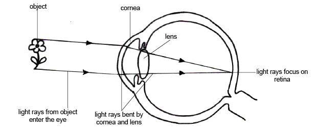
The eye is a fairly simple sensory organ which functions in the following way:
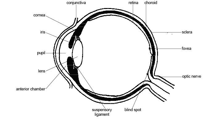
Light reflected off the observed object, enters the eye and is focused by the lens on the retina at the back of the eye. This stimulus is sent to the brain via the optic nerve where it is interpreted as a visual image. The eye has the following basic structure:
The lens is a transparent, convex structure made of connective tissue which focuses the light entering the eye on the photo-sensitive retina at the back of the eye. The lens is held in place by suspensory ligaments (also known as zonules) just behind the pupil which is a hole in the coloured part of the eye known as the iris. In front of the lens, iris and pupil is an anterior chamber or space filled with a watery substance known as the aqueous humour. Behind the lens is a posterior chamber or space filled with a more jelly-like substance known as the vitreous humour. These liquids exert a hydrostatic pressure inside the eye enabling it to maintain its spherical shape. Lens luxation is the name give to a movement of the lens into an incorrect position in the eye. This is caused by deterioration or damage to the zonulesso that they are unable to hold the lens properly or in the correct position. If the lens is still held by some zonules and has moved just a little out of position it is said to be subluxated. If the lens is no longer held by any zonules it is fully luxated. Lens luxation is called Primary Lens Luxation if it is the first thing to go wrong with the eye. Primary Lens Luxation can either be caused by degeneration and breakage of the zonules which are supposed to hold it in place or by an injury to the eye which damages the zonules. Another eye affliction, for example infection, inflammation, a tumour or glaucoma, can damage the zonules and cause the lens to move out of position, which is known as Secondary Lens Luxation. In the case of predisposed breeds of dog, the Primary Lens Luxation is an inherited condition whereby the zonules degenerate and break causing luxation. This Primary Lens Luxation can cause glaucoma as a secondary problem which is very serious condition which results in blindness. Many breeds of dog are affected by PLL e.g. Jack Russell and Parson Russell Terriers, Fox Terriers, Scottish Terriers, Welsh Terriers, Sealyham Terriers, Skye Terriers, Manchester Terriers, Tibetan Terriers, Cardigan Welsh Corgis, Lancashire Heelers, Border Collies, Australian Cattle Dogs, Brittany Spaniels and of course, Miniature Bull Terriers. PLL in these breeds is believed to be caused by a gene mutation. Carriers of the gene may suffer from the condition or they may not, but they all can pass it on to their offspring. The gene causes the zonules to degenerate and break so that they no longer can hold the lens in the correct position. The lens may move forward into the anterior chamber, in which case, if treatment is sought quickly it can be surgically removed. This procedure, if done in time, can save some sight in the eye for the animal. The lens may move backward into the posterior chamber, in which case, it is very difficult to surgically remove it. If the displacement is not too severe and it is still near to the iris, medical eye drops can be used to maintain a small enough pupil to trap the lens close to the iris. If the lens moves too far into the back of the eye there is risk of it damaging the retina. Both anterior and posterior luxation can cause glaucoma which is increased pressure in the eye. Glaucoma is an extremely serious condition which if left untreated for even as little at 6 hours can result in permanent damage to the retina of the eye and the complete loss of sight. Interestingly if glaucoma can be avoided, dogs that have had both lenses removed can still see well enough to live normal lives. The cornea is able to focus light sufficiently for the brain to compensate and interpret such that in some cases the animal appears to suffer no loss of sight at all. The symptoms of PLL are pain (seen by rubbing or pawing at the eye which can also cause redness and swelling), tearing or watering of the eye and loss of sight. Sometimes, the eye can appear asymmetrical or cloudy. Any change in eye appearance should be followed up immediately with a veterinarian. PLL should be considered a medical emergency as treatment in the first 24 to 48 hours can prevent the damage that can cause permanent blindness. Unfortunately if a dog of a predisposed breed develops PLL in one eye, it is almost certain that the other eye will also become affected soon afterwards. Inherited PLL tends to appear only at the ages of 4 to 7 years. This is a big problem, as in most cases, these animals have already been bred and produced offspring. After many years of research, In September 2009, the Animal Health Trust identified the gene mutation causing this problem and design a genetic test for it to identify carriers before they produce any offspring. A cheek swab sample is taken and the possible results of the DNA test are: PLL CLEAR - this dog has two copies of the normal gene and will not develop Primary Lens Luxation as a result of the mutation being tested for. PLL CARRIER - this dog has one copy of the mutation and one normal copy of DNA, is unlikely to develop Primary Lens Luxation but may pass the mutation onto their offspring. PLL AFFECTED - this dog has two copies of the mutation and is at risk of developing Primary Lens Luxation. This dog will pass on the mutation to all its offspring. PLL is almost unknown in Standard Bull Terriers.
It is essential to DNA test all Miniature Bull Terriers before
breeding to ensure that no PLL-affected dogs are produced and
to responsibly manage and eliminate the mutation over time and
protect our breed from this genetic threat. |
|
Home | History | Imports | Litters | Profiles | Exports | In Memory | Interest | Archives | Links Copyright © 2009 - 2011 Tracey Butchart - Miniature Bull Terrier Network - South Africa - All Rights Reserved |